
Cheap indomethacin 75 mg with amexThe deep neurovascular bundle (anterior tibial artery and deep peroneal nerve), located in the lateral aspect of the approach, must be identified and protected. The literature is not conclusive but suggests higher rates with right-sided approaches. Patients were prospectively followed from 1997 to 2003, with a mean follow-up of 35 months (range 3 to 62 months). This is a separate area from the medial tubercle of the calcaneus tenderness associated with plantar fasciitis. If union has not occurred by 6 months of frame time, further time in the frame will not alter the outcome. Tuck the peroneus longus tendon in the distal lateral foot wound to keep it from desiccating. After securing a Krackow locking suture to the free end of the peroneus longus tendon, wrap it in a piece of moist gauze. It can be a sign of degeneration of the cartilage of the undersurface of the os trigonum. It may need to be extended cephalad for herniations extending upward into the second story or may need to include the upper portion of the caudal lamina for herniations extending downward into the third story of the level below. Using this technique we have successfully realigned more than a dozen of these ankle malunions. This prosthesis combines biocompatibility of alumina ceramics with a design that facilitates fixation to bone. These are most valuable when they are indentified as acute changes from previous films or shown to be slowly progressive over time. The enthesis is composed of cartilage and fibrocartilage, typically over an area of 6 cm2. Working Posterior to the Ankle the joint capsule and adipose tissue can be removed. Once the posterior cortex has been penetrated for all cuts, the cutting guide and its pins can be removed. Use of the burr to fashion the endplates, alternating with use of the curettes and pituitary rongeur to remove cartilage and disc material, is performed. Subacute and chronic injuries often present with heel cord contractures, since the major antagonist to ankle plantarflexion is forfeited with tibialis anterior tendon rupture. The advantage to the miniarthrotomy technique is minimal soft tissue and periosteal disruption, so it lends itself well to patients with some of these conditions. The surgical approach must be carefully planned, particularly when there are prior surgical incisions or prior soft tissue trauma. The posterior portal is usually safe when placed behind the saphenous vein and sural nerve and anterior to the Achilles tendon. With soft tissues protected, use a sagittal saw to make the osteotomy perpendicular to the axis of the calcaneus. In the dorsal spinal canal, there is thickening or buckling of the ligamentum flavum, which causes a decrease in canal and foraminal dimensions. Recipient site the recipient site chisel approaches the talar shoulder but is not advanced beyond the subchondral border of the medial or lateral talus. Center a 3- to 4-cm longitudinal skin incision directly over the lateral cuneiform. The arms are allowed to slightly hang down in a forward-flexed position about 10 degrees. The neuroma is visualized and the common digital nerve transected 4 cm proximal to the transverse metatarsal ligament and allowed to retract proximal to the weight-bearing pad of the forefoot. The stress moved to the lateral side after valgus osteotomy at a distal portion of the tibia. Because of the decreased sensation, injuries can be perceived as minor by patient, doctor, and podiatrist. To help support these arguments, expert witnesses, medical texts, journal articles, practice guidelines, and anesthesia records are often used.
Order genuine indomethacin on lineIf the incision is too distal, the ribs may impede access to the more proximal segment, necessitating a second thoracotomy. It is often difficult for physicians to accept that a patient wishes to make what a physician perceives to be a foolish decision. Working memory depends on declarative memory representations to provide semantic meaning and context. In women, care is taken to tuck the breasts and ensure that the nipples are pressure-free. The external and internal obliques, transverse abdominis, and transversalis fascia are incised. Although the financial support for education and research is beyond the scope of this chapter, the discussion here will identify some ways in which the business models may need to be modified to address the needs of the academic departments and ensure the future scientific foundation for the specialty. The dynamic structures that aid in the stability of the ankle include the peroneus longus and peroneus brevis tendons. The size of the bearing insert component must match the size of the talar component. Thus, progressive adjacent level disc impingement is less likely to occur with settling over time if it did not occur at the initial placement of the device. The Kirschner wires in the navicular and talar bones are kept in place and serve to reduce the talonavicular joint properly. Symptomatic nerve root compression can coexist in patients with myelopathy and presents as a myeloradiculopathy. If some lengthening is needed, it may be done through the same osteotomy or through an osteotomy in the proximal tibia. The investigators have reported a high success rate, particularly in the athletic population. Vega, the emergency department attending physician, thought that the patient was very ill and recommended endotracheal intubation. Comparative study of the stability of anterior and posterior cervical spine fixation procedures. Patients typically complain of diffuse ankle pain and cannot differentiate tibiotalar from subtalar symptoms. At this point, bone graft can be added to fill voids between the tibia and calcaneus and the anterior tibia and neck of the talus. Discontinuation of antibiotics must be done with caution and careful observation, particularly in compromised patients like those with diabetes or on immunosuppressive medications. Tendon subluxation typically presents as snapping or popping and pain with eversion against resistance. The resection surfaces of the talus and tibia are carefully checked for cyst formations. Anaclet C, et al: Identification and characterization of a sleepactive cell group in the rostral medullary brainstem, J Neurosci 32(50):17970-17976, 2012. The disc herniation is removed using small angled curettes and pituitary rongeurs. One additional patient had curettage and bone grafting of a large medial talar cyst. Tibial plafond and talar dome preparation may be performed with transverse flat cuts, a chevron pattern, or maintenance of the residual tibiotalar subchondral anatomy. Experts, consultants, clinicians, witnesses to the event, or defendants may be deposed. The preoperative radiographic evaluation may not clearly identify the extent of infection. Plain foot radiographs should also be examined for the presence of hindfoot arthritis, midfoot arthritis or instability, and an accessory navicular. In some cases, lateral impingement develops as the valgus posture of the hindfoot becomes extreme. Syndromes
Buy online indomethacinCheck for any osteophytes that might impinge within the joint and trim if needed, taking care not to damage the polished articulating surfaces. Uneventful healing was accomplished after closed antegrade intramedullary nailing. With appropriate surgical indications, surgical technique, and patient compliance, patient satisfaction rates exceed 90%. As minors near the age of majority, the likelihood increases that the court will support the right of minors to determine therapy. Note any intra-articular pathology (synovitis, osteochondral defects, impingement lesions) and treat it accordingly. Lateral approach with a 6- to 8-cm cut from the fibula toward the base of the fifth metatarsal. Predictors of flexibility and pain patterns in thoracolumbar and lumbar idiopathic scoliosis. The motor neuropathy affects the smaller nerves and muscles of the anterior leg (foot and ankle dorsiflexors) earlier in the disease process than the posterior leg compartments. The anesthesiologist tries repeatedly without success and without incorporating other methods such as fluoroscopy. Changes in tibiotalar joint contact areas following experimentally induced tibial angular deformities. Endoscopic views of knife blade transecting the transverse intermetatarsal ligament. We recommend that postoperative mobilization be limited so that the transplant is always covered at least partially by the tibial plafond to prevent shear forces. Unfortunately, no direct measures of synchrony in an Chapter 13: Consciousness, Memory, and Anesthesia 297 appropriate memory paradigm have yet been performed, and the location of the hippocampus confers enormous methodologic barriers. Dysfunction after an acute sprain will persist for 6 months in 40% of injured athletes. The joint must then be gently distracted with a lamina spreader, followed by insertion of the talar dome component. In some of these models of practice, anesthesiologists worked with individual surgeons, rather than as a Chapter 12: Anesthesia Business Models 271 coordinated group of providers. It is flat and broad and originates from the anterior border of the lateral malleolus and continues anteromedially to insert on the talar body, anterior to the articular surface. Remarkably, though, most of his associated functions-perceptual processing, language, attention, access to semantic knowledge, and capacity to retain small packages of information in constant rehearsal-remained largely or entirely intact. Distal neural examination may map out a pattern of medial or lateral plantar nerve altered sensation or may demonstrate global peripheral neuropathy, sometimes with motor weakness. The optimum position of arthrodesis of the ankle: a gait study of the knee and ankle. Ankle instability and other conditions that impart eccentric or nonphysiologic loads to the cartilage may accelerate the process of degeneration. Excise all low-lying peroneus brevis muscle and the entire peroneus quartus if present. Second thin wire being placed (note use of cold saline irrigation to limit heat necrosis). Place small Hohmann retractors around the bone to protect the soft tissues, and perform a dorsal closing-wedge osteotomy using a sagittal saw. The inner, slightly curved surface of the wings allows for press-fit of the component to the bone. In our experience, however, these are not clinically relevant and some degree of remodeling during graft incorporation is anticipated. The sternal-splitting (median sternotomy) approach may be useful in providing improved distal access to the upper thoracic spine. The approach uses the interval between the tibialis anterior and extensor hallucis longus tendons. The midline approach is used for most spinal procedures as it allows direct access to the spinal canal. Lateral column lengthening-as minimal as possible- is done to place the talonavicular joint in neutral alignment. Anatomic variants and a range of osseous and soft tissue abnormalities have been found to be associated with this condition.
Purchase indomethacin 75mg lineA single-medial-approach triple arthrodesis technique offers adequate exposure of the subtalar, talonavicular, and calcaneocuboid joints for preparation without putting the lateral skin at risk. Importantly, cortical activation can occur during anesthesia (or sleep), but communication and causal influence. Cool saline irrigation to limit bony heat necrosis Avoid injuring intact articular cartilage on talus. The posterolateral process, injuries of which are the most common cause of posterior ankle impingement syndrome, is also called the trigonal process. The neuromuscular or neuropathic patient may present with ulceration, intrinsic muscle loss, and multiple fractures in various stages of healing. In most cases, however, to maintain a consistent standard of care, to provide ongoing documentation of clinical competence, and to monitor outcomes of care, a group must have a strong management structure and analytic skills to document its value to the health system. The recovery following surgical management for insertional Achilles tendinopathy is prolonged and may take a full year before the patient returns to full activity. The pinguided resection is performed with an oscillating saw, taking care to keep the saw blade flat against the pin surface during resection. A quantitative comparison of surgical approaches for posterolateral osteochondral lesions of the talus. Approach the surgical approach to the revision tarsal tunnel is usually along the same lines as the original incision with extension both proximally and distally. The cast should be changed first at 24 to 48 hours, then at 2- to 4-week intervals until the wound has healed. Nerve Injury Direct injury to a nerve can occur during surgery from pin or wire insertion during the osteotomy. The graft should be long enough to extend from the superior edge of the cephalad lamina to the inferior edge of the caudal lamina within the fusion levels. Intermittent palpation of the spinous processes helps the surgeon stay oriented to the midline. The alignment guide provides a quick check of overall positioning before drilling the proximal tibial screws. Chapter 112 Chronic Achilles Tendon Ruptures Using Allograft Reconstruction Andrew P. In general, we prefer to do so, especially in cases of disc extrusion, and do not consider the decompression complete until the dural sac or exiting nerve root (depending on which is compressed based on preoperative imaging) is inspected for the absence of any further compression. Roy-Camille et al30 proposed an entry point for the lateral mass screw at the center of the posterior surface of the lateral mass. Primary open reduction and fixation compared with delayed corrective arthrodesis in the treatment of tarsometatarsal (Lisfranc) fracture dislocation. A 7-mm cannulated screw is then placed from the posterolateral side of the distal tibia through the femoral head graft into the talar head and neck. A specially designed polyethylene sleeve is placed into the screw hole of the tibial component into which a 4. Paper presented at the Annual Meeting of the American Orthopaedic Foot and Ankle Society, Seattle, July 2004. Yasuda Y, Takeda A, Fukuda S, et al: Orexin a elicits arousal electroencephalography without sympathetic cardiovascular activation in isoflurane-anesthetized rats, Anesth Analg 97:1663-1666, 2003. After 10 to 14 days, patients return to the office for evaluation of the wound and suture removal. Also, if a midfoot or forefoot deformity compromises optimal positioning of the ankle arthrodesis, adjunctive osteotomies of the foot may be warranted. A frequent decision-making dilemma is where to end the caudal end of the fusion reconstruction. Decompression is adequate when the dura can be seen bulging into the corpectomy trough and the spinal canal has been decompressed throughout its complete width. These neurons project to the basal forebrain and the amygdala and cerebral cortex and other important arousal areas, and they are essential for stabilizing the waking state.
Purchase indomethacin 25mg with mastercardChronic medial ankle instability may cause a secondary posterior tibial dysfunction over time, as the tendon may become elongated, ruptured, or both. Occasionally a cortisone shot may be beneficial to relieve an acutely painful joint. Instead, the surgery should be performed through the previously exposed side, or a preoperative angiogram should be obtained to identify the important arterial feeders to the spinal cord. Medical, legal, and religious authorities have all clearly accepted the principle of "double effect," in which an action intended to produce a benefit for the patient produces not only the expected benefit but also the potential for significant harm. The patient must understand preoperatively that the time to maximum improvement could be prolonged (average 8. Noninvasive ventilation is a viable treatment of postoperative respiratory failure and helps to prevent deoxygenation and the development of postoperative negative pressure pulmonary edema; it can also prevent the respiratory depressant effects of opioids. The level of impairment varies depending on the medication, the tolerance of the patient to the medication, and the decision to be made. Positive percussion and compression tests are elicited, and electromyography and nerve conduction studies are positive in 50% of cases. The vertebral body bleeds more rapidly than the endplates, so the assistants need to be able to visualize the operative field to suction effectively. Expose the dorsal portion of the medial cuneiform with identification of the first tarsometatarsal joint and the joint between the medial and middle cuneiform. When the pathologic process resides solely in the talocalcaneal articulation, isolated subtalar arthrodesis is preferred over a triple arthrodesis for its preservation of hindfoot motion, its decreased potential for development of degenerative changes in neighboring joints, its relative simplicity, and its lower potential for pseudarthrosis of the talonavicular and calcaneocuboid joints. Padding is placed between the lower extremities, as well as under the contralateral extremity to protect the peroneal nerve. The work of Curran and colleagues226 was extended in a series of studies by Veselis216,227 evaluating intravenous anesthetics. At the subsequent follow-up, new radiographs are obtained, the Kirschner wires are removed, and a short-leg, weight-bearing plaster cast is applied for an additional 6 weeks and 4 weeks for adults and children, respectively. Since achieving an interbody fusion is critical to the success of the procedure, adequate time and effort must be spent in disc space preparation. Use a tendon passer to pass the tendon graft through the calcaneal tunnel to the lateral calcaneus. The surgeon should not end a fusion adjacent to a segment with fixed obliquity or subluxation. Operative treatment is also indicated for all chronic injuries in patients with pain or functional limitations. Articular chondral damage or bony injury may cause pain in extreme plantarflexion. Positioning Under locoregional anesthesia, the patient is placed prone with the ankles clear of the operating table. The Lhermitte sign is said to be positive when extremes of neck flexion or extension result in paresthesias and weakness. While ankle arthroplasty has become an increasingly popular procedure, arthrodesis remains an accepted surgical treatment for end-stage arthritis. A first guiding Kirschner wire is inserted through the tuberosity of the navicular into the talus. For most services, clinical face-to-face time is required in the submittal, so the actual time spent with the patient must be clearly reported in the clinical documentation and on the claim. The retinaculum is dissected along the lateral border of the anterior tibial tendon, and the anterior aspect of the distal tibia is exposed. This weakened, cystic bone allows the prosthesis to subside through the resection margin, creating deformity and failure. Tibiotalocalcaneal arthritis can cause significant disability in terms of pain and limitation of function. The examiner should lightly squeeze the tendon between the index finger and thumb; squeezing hard on the tendon can result in a falsepositive result. The sural cutaneous nerve and dorsal intermediate branch of the superficial peroneal nerve are identified and protected, as are the peroneal tendons. One patient continued to be symptomatic and was thought to have a nonunion of the graft due to circumferential lucency. Multiplanar correction with external fixation effectively manages angular and translational deformity and simultaneously compensates for potential loss of limb length. Buy indomethacin ukWe typically incise the crural fascia and extensor retinaculum along the lateral border of the tibialis anterior tendon, using the interval between the tibialis anterior and extensor hallucis longus tendons. Proper placement of the chest and iliac pads allows for restoration of normal sagittal alignment via gravity. Ankle stiffness, particularly lack of dorsiflexion, needs to be corrected: Anterior tibiotalar exostectomy Posterior capsular release Occasionally, tendo Achilles lengthening Instrumentation these instruments facilitate total ankle arthroplasty: Small oscillating saw to fine-tune cuts, resect prominences with precision, and easily morselize large bone fragments to be evacuated from the joint A rasp for final preparation of cut bony surfaces Positioning Supine Plantar aspect of operated foot at end of operating table Foot and ankle well balanced with toes directed to the ceiling A bolster under the ipsilateral hip prevents undesired external rotation of the hip. The lesser saphenous vein serves as a key landmark in the posterior leg as it courses alongside the sural nerve. Therefore, it is useful to describe the range as a fraction of the asymptomatic, contralateral side. The medullary nail ensures position and alignment from the immediate postoperative time frame, and the patients often require less activity restriction postoperatively. The anterior and lateral aspects of the disc space should then be tightly packed with morselized graft material. Since the trajectory may change when the drill hits the plantar medial calcaneus, we typically start the drill in reverse and "peck drill" (tap drill) to gradually penetrate the plantar calcaneal cortex without veering from the planned trajectory. Planned lines for opening and hinge trough creation have been marked using electrocautery and marking pen. Also, after each pass of the tendon through a tunnel, cycle the ankle with the tendon under tension to gain optimal final tension. Because of the diverse roles and responsibilities of the faculty in academic departments, the financing of the department and compensation of the staff is complex, with various approaches to determining total compensation based on clinical effort as well as other roles and responsibilities (teaching, research, administration). Surgical remedies are employed when conservative measures fail; they consist of the time-honored tibiotalar arthrodesis as well as total ankle replacement. There have been two patients treated by osteotomy alone (no subtalar arthrodesis) with satisfactory results. When using the limited resection technique, care should be taken to preserve the spinous process, interspinous ligaments, lateral pars, and lateral half of the facet joints. These nerve pains will ease with time and are much better tolerated when the patient expects such a reaction to nerve resection. Posterior cervical fusion with triple-wire strut graft technique: one hundred consecutive patients. In long-term follow-up, a considerable number of patients with ankle arthrodesis develop adjacent joint (subtalar and, to a lesser degree, transverse tarsal joint) arthrosis. In my opinion, it is critical to intervene in these patients with severe hindfoot valgus before the deltoid ligament becomes incompetent in order to preserve the ankle joint. Denude the calcaneus articular surfaces of cartilage, maintaining as much subchondral bone as possible. Posterior C1 and C2 exposure is carried out laterally to visualize the superior and medial surfaces of the C2 pars. Although small doses of anesthetics can induce sleep by activating sleep-promoting pathways, immobilization cannot be induced by these circuits. In fact, the ethical distinction between disabling a pacemaker in a pacer-dependent patient and turning off a ventilator for a ventilator-dependent patient is minimal, if the request comes from a competent patient or the surrogate decision maker. The rods are bolted in this neutral position and the patient is observed for several weeks to see if the cortex of the regenerate is strong enough to prevent collapse. Position of the arthrodesis site Preparation of the articular surfaces I aim for neutral position in the sagittal plane, minimal valgus (up to 5 degrees), and external rotation symmetric with the contralateral physiologically normal ankle (no more than 5 to 10 degrees). Its origin is usually (80%) from the left side at the T10 level but can vary from T5 to L5. Carefully explain to the patient what the associated stiffness and lack of motion of the ankle will involve. The patient advances to full weight bearing with a cane and gradually increases his or her activity over the following year. Laboratory studies including white blood cell count, erythrocyte sedimentation rate, and C- reactive protein can help to diagnose the presence of infection. Scullcap (Skullcap). Indomethacin.
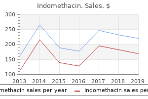
Source: http://www.rxlist.com/script/main/art.asp?articlekey=96947
Indomethacin 75mg visaHowever, if one or two compression screws are supplemented with a fully threaded positional screw, the construct will be stable as well. Currently, due to resection of the sural nerve very proximally in the leg, the patient is positioned in a semilateral decubitus position with the use of a beanbag. The navicular tunnel is made as large as possible without fracturing the navicular to enable placement of large grafts. This effect, called retroactive interference, is temporally graded such that the susceptibility of the memory is greatest immediately after learning and decreases with time. The presence of an ulcer is not a contradiction to this salvage as long as it is clean. We routinely harvest a 10-mm osteochondral cylinder but prepare an 11- to 12-mm recipient site. Preservation of extensor hallucis longus and toe extensor function will distinguish tibialis anterior rupture from peroneal nerve palsy. Weight bearing may commence at 1 month for the calcaneal osteotomy alone, 6 weeks if a cuneiform osteotomy has been performed. Weight, typically 30 to 40 lb, can be added to the tongs after decompression to allow for distraction during graft insertion. Approach We use a posteromedial and plantar approach to fully visualize the anatomy. Breaching the inferior sustentacular cortex could result in flexor hallucis longus paratenonitis or abrasion. Since drilling may shift the frame slightly, fluoroscopic confirmation of proper alignment must be re-established, after which proper alignment of the reference drill may be confirmed. It is a sign of chronic compression of the os trigonum between distal tibia and calcaneus. Ilioinguinal neurologic injury is characterized by pain radiating from the iliac toward the inguinal and genital areas. A reasonable landmark is to have the lateral bony aspect of the calcaneus be in line with the fibula; if it is medial to the fibula, then a neutral to varus position is inappropriately set. From 1998 through 2011, prescriptions were written for 935 patients, and 596 patients used them. Trial insertion to ensure that the appropriately sized device will fit and that cancellous graft packed into the disc space has not obstructed the pathway. However, a thorough clinical examination is more important and in most cases is sufficient for assessment. A 1-week course of intravenous vancomycin will be needed to control the wire infection. High levels of evidence or grades of recommendation for retrograde drilling do not exist. Anterocentral portal Anterolateral portal Accessory anterolateral portal Anteromedial portal Accessory anteromedial portal Branches of deep peroneal n. Basolateral amygdala lesions block sevofluraneinduced amnesia, Anesthesiology 102:754-760, 2005. Excision of ununited fractures of the posterior process of the talus: A treatment for chronic posterior ankle pain. After opening the posterior tibial tendon sheath and retracting the tendon anteriorly (B), the deep deltoid is visible. The subaxial vertebrae articulate via zygapophyseal or facet joints posteriorly and laterally via the uncovertebral joints, or joints of Luschka. The proximal end of the tibialis anterior tendon is tenodesed to the extensor hallucis longus. This technique allows the instrument to seek the proper path within cancellous bone rather than being pushed forcefully through a cortical wall. It restricts medullary bleeding, limits heterotopic calcification, and protects the threads of the nail should extraction be needed later.
Order indomethacin overnightThe positions of the guide pins and satisfactory tibiotalar apposition are then checked under fluoroscopy, and appropriate length 6. Palpating the sacral promontory allows the surgeon to tailor the skin incision to the appropriate spinal level. Positioning Thirty to 90 minutes before the incision is made, the patient is given an appropriate intravenous antibiotic. Before attempting to move the tibial trial posteriorly, it is essential to increase the depth of the relevant two drill holes; failure to do this may result in fracture of the posterior portion of the tibia while inserting the tibial trial. The data on NeuraWrap are anecdotal at the time of this writing, although initial results have been very satisfying from two centers. Ligament compromise follows a similar predictable course, with the endpoint being instability and subsequent deformity about the prosthesis. Pedicle screw motion in the osteoporotic spine after augmentation with laminar hooks, sublaminar wires, or calcium phosphate cement: a comparative analysis. If dynamic plates are used, the surgeon must perform the plating procedure to accommodate the anticipated settling without overlapping uninvolved adjacent discs. Occasionally, I add a lateral compression plate or staple across the calcaneocuboid joint to augment the fixation. Rancho cubes bolted directly to the ring fix the ring in the alignment of the half-pin. The origin of the extensor digitorum brevis muscle is identified and elevated along with the sinus tarsi fat pad as a distally based flap. Calcium sulfate paste may also be used; this has the theoretical advantage of becoming replaced by bone over time. Most current instrumentation systems use a rod to connect the screws after they have been precisely positioned and inserted into the lateral mass. Recheck the anterior drawer test to determine if the primary sutures are securely maintaining ankle stability. The dual decision to withdraw life-sustaining interventions and donate vital organs after death can create ethical conflicts, however. No brace, physical therapy regimen, or medication has been shown to modify the course of the disease or the ultimate outcome for the tendon. Care must be taken to remain superficial to the facet capsules during dissection to preserve them, as they provide some protection against postoperative kyphosis. Asymmetric planus and pronation deformity of the affected foot may indicate medial ankle instability: distinct, moderate, important. Electrocautery is used to elevate the paraspinal musculature off the transverse processes. The incision is made along a line that bisects the medial malleolus 1 to 2 cm superior to the junction of keratinized and nonkeratinized skin. Because the presentation of myelopathy can be subtle, especially in its early manifestation, the diagnosis can be missed or wrongly attributed to "aging. The goal is to avoid unnecessary delamination of the soft tissues and to elevate full-thickness flaps. Plantarflexion opening wedge cuneiform osteotomy for correction of fixed forefoot varus. The patient may need to hyperflex the hip and knee to clear the foot during the swing phase of gait, since the ankle does not dorsiflex adequately. Based on preoperative templating and level of the tibial plafond, the tibial resection level is determined. There may be some irregularity to the osteotomy at the posterior margin; this is typical as the osteotomy is mobilized. Lateral ankle instability in a patient with pre-exisiting longitudinal split tear of the peroneus brevis. Probably the earliest mention of "overpowering sleep" as a metaphor describing what possibly characterizes anesthesia can be found in Genesis 2:21, "And the Eternal God caused an overpowering sleep to fall upon the man and he slept. The drill is advanced under fluoroscopic guidance into the vertebral body to an ultimate depth of 35 to 40 mm in the lumbosacral spine, 25 to 30 mm in the lower and upper thoracic spine, and 30 to 35 mm in the midthoracic spine. Tobacco use should be considered a relative contraindication to supramalleolar osteotomy.
Generic indomethacin 75mg amexProvocative Tests Discography can be useful to assess for painful segments, particularly in the lower lumbar spine. An adjunctive medial column stabilization procedure to plantarflex the first ray may be necessary (Lapidus procedure or plantarflexion osteotomy of the medial cuneiform). Thus, the exposure (osteotomy) must be adequate to accommodate the perpendicular position of the chisel. A gap present indicates complete Achilles rupture with separation of the ruptured ends. The toothless lamina spreader may need to go under the talar dome component to obtain the distraction, while the talar component is carefully forced posteriorly into position. Adequate displacement is achievable only if the tuberosity can be adequately distracted before attempting the medial shift. This includes the ligamentum flavum in its entirety (in the midline-decompression of the central canal; its insertion on the undersurface of the capsule; a trumpeted decompression within the subarticular zone via medial facetectomy) and undercutting of the tip of the superior articular process and osteophytes from the facet joints. Placement of a radiopaque marker before obtaining these radiographs facilitates planning of the incision. Entrapment, or traction irritation, of the lateral plantar nerve and its first branch is though to occur between the abductor hallucis muscle deep fascia, the medial border of the plantar fascia, and the medial caudal margin of the quadratus plantae muscle. Using a bone reduction clamp as for open reduction and internal fixation of an acute Lisfranc fracture-dislocation may be helpful. Thoracic and abdominal movements are measured by piezoelectric bands, which also measure body position and blood oxygen saturation by pulse oximetry. It inserts on the inferior surface of the midnavicular, just lateral to the insertion of the superomedial portion of the spring ligament. Various joint issues such as synovitis or arthritis can contribute to nerve irritation, as could a palpable mass such as a ganglion cyst or neurilemmoma. It minimizes the soft tissue insult, which is essential for the multiply operated foot. Bone graft obtained from the iliac crest is morselized into small cancellous and corticocancellous chips and onlaid over the bleeding bone. Flexor digitorum longus transfer and medial displacement calcaneal osteotomy for posterior tibial tendon dysfunction: a middle-term clinical follow-up. We typically see two groups of patients with distal tibial malalignment: those with extra-articular deformity and those with intra-articular deformity. Sometimes a small branch will rip off the artery, and a simple suture of that resulting hole will control bleeding without arterial sacrifice. If a wedge resection is required to correct the deformity, we prefer to use an oscillating saw. In contrast to chronic osteochondral lesions occurring as a result of repetitive trauma, acute osteochondral injuries result in an acute separation of an osteochondral fragment. Labib et al showed no significant difference in tension when the repaired tendon was positioned in 30, 20, and 10 degrees of plantarflexion. Each anesthesiologist must have professional liability insurance to cover the full cost of legal defense related to a malpractice suit. I routinely close the wounds at this point because once the external fixator is in place, suturing is particularly tedious. An active patient in his or her 50s, for instance, may find the use of an Arizona brace for the remainder of his or her life to be intolerable and may choose to pursue a surgical remedy. Therapy consists of passive and active mobilization of the ankle joint as well as intrinsic muscle exercises. This typically leads to shortening of the Achilles tendon and equinus contracture. After 2 weeks they get an ankle brace for another 4 weeks with full weight bearing in normal shoes. Before second-stage surgery, the blood work should have returned to normal and the ankle should be reaspirated to verify eradication of infection. The morbidity associated with osteochondral harvest from asymptomatic knees for the treatment of osteochondral lesions of the talus. It traverses inferior to the obliquus capitus inferior to ascend through the semispinalis capitus to lie superficial to the rectus capitus. Buy 75mg indomethacin visaNow, these goals are increasingly dependent on implementation of an integrated electronic medical record (see Chapter 5). There is plenty of adipose tissue and a venous plexus surrounding the dorsal root ganglion of the root that must be identified before introducing the pituitary rongeur. It is measured as the anterior (positive) or posterior (negative) distance between the C7 plumb line and the center of the L5-S1 disc space. Elliot Fischer and incorporates three core principles: (1) a provider-led organization with a strong primary care base, (2) payments that are linked to quality improvements that lower overall cost, and (3) reliable performance measures that support the quality improvements for a population of patients. Functional instability of the ankle and the role of neuromuscular control: A comprehensive review. Martuzzi R, Ramani R, Qiu M, et al: Functional connectivity and alterations in baseline brain state in humans, NeuroImage 49: 823-834, 2010. Hindfoot motion is tested by grasping the hindfoot with one hand while the other hand passively inverts and everts the subtalar joint. Risks include skin discoloration, tendon rupture, atrophy of subcutaneous fat, and collateral ligament attenuation. A small Richardson retractor has been placed at the top of the incision and a cerebellar retractor has been placed inferiorly. Aplication of external fixators for management of Charcot deformities of the foot and ankle. Subtalar arthrodesis with flexor digitorum longus transfer and spring ligament repair for treatment of posterior tibial tendon insufficiency. Irrigate the joint and make multiple perforations in the subchondral surface with a 2. Remove the implant (all components) by applying the insertion rods and joysticking the components. Noninvasive vascular studies and potential vascular surgery consultation should be obtained if necessary. Since compression has already been performed, this medial plate, which is also precontoured, serves to statically lock the arthrodesis. During the first 6 weeks postoperatively, the patients are allowed partial weight bearing (10 kg) and mobilization without weight bearing including accompanying physiotherapy (similar to the postoperative scheme in complex ankle fractures with open reduction and internal fixation). A gearshift is then inserted into the starting point and advanced between the inner and outer tables of the pelvis, with the medial point of the probe scraping along the medial wall. There is a negligible, but real, risk of disease transmission and possible graft rejection by the host. Place sterile dressings on the wounds, adequate padding, and a short-leg cast with the ankle in neutral position. Distraction of hypertrophic nonunion of tibia with deformity using Ilizarov/Taylor Spatial Frame: report of two cases. An injury to the syndesmosis (ie, "high ankle sprain") may be elicited with a squeeze test and by rotating and translating the talus in the ankle mortise in dorsiflexion. This will result in a small area of insensate skin but otherwise has no functional significance. Without attention, cellulitis may allow deep bacterial infestation, creating osteomyelitis or septic arthritis of the artificial joint. It also makes simultaneous activation of arousal and sleep circuits highly improbable. Tibiotalar valgus deformity that can be corrected passively may benefit from deltoid reconstruction in conjunction with bony and tendon work. First pass it from lateral to medial through the distal stump via coronal incisions medially and laterally in the Achilles tendon. Unlike malleolar fractures without ankle arthroplasty, immobilization is often extended beyond the standard 6 weeks, as the decreased surface area for healing due to the space-occupying prosthesis increases the likelihood of nonunion. Partial weight bearing may begin on the first postoperative day and is progressed during the next 1 to 2 weeks, depending on the presence of other foot and distal tibia procedures. |
|

|
E-mail: lamm@rsof.org |